Mapping how the brain organizes movement
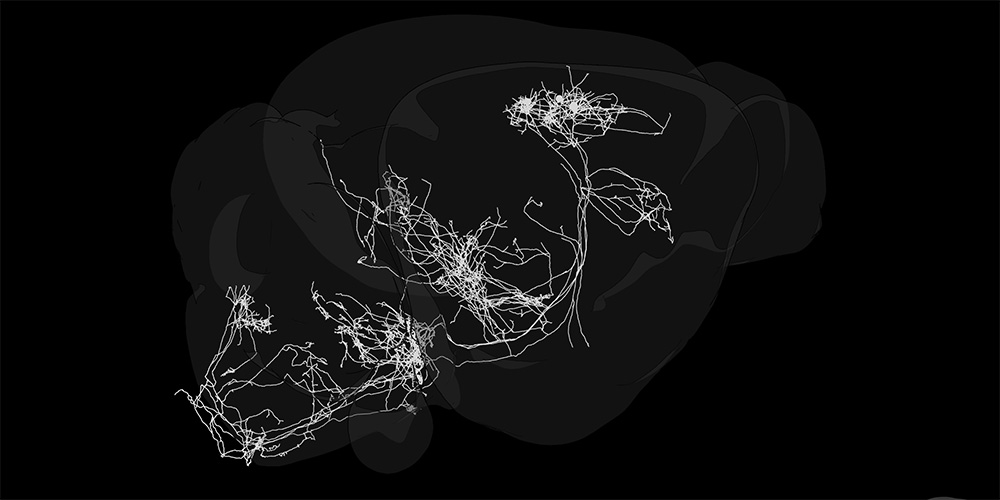
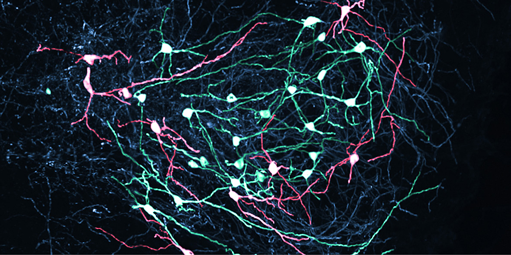
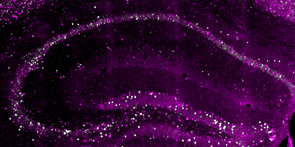
Researchers at the Biozentrum of the University of Basel and the FMI have created the most detailed map yet of the mouse motor cortex. The map reveals 16 distinct subregions and sheds new light on the brain circuits that control movement. The findings could help researchers better understand conditions that affect these circuits, including amyotrophic lateral sclerosis and dementia.
24 September 2026
Every movement – from reaching for a cup to turning our head – depends on the brain combining information about the body, surroundings and the actions we want to perform. But how some key brain regions that control movement are organized remains unclear, including the motor cortex.
In mice, standard brain atlases typically divide the motor cortex into broad primary and secondary regions, based on differences in connectivity. A new study from Professor Silvia Arber’s team at the FMI and the Biozentrum of the University of Basel, together with collaborators at the Allen Institute, reveals a much finer level of organization.
The researchers identified 16 distinct subregions, each defined by its own pattern of connections with other parts of the brain. The map provides a precise framework for studying how movement is controlled and how motor-cortex circuits may be affected in disease.
-
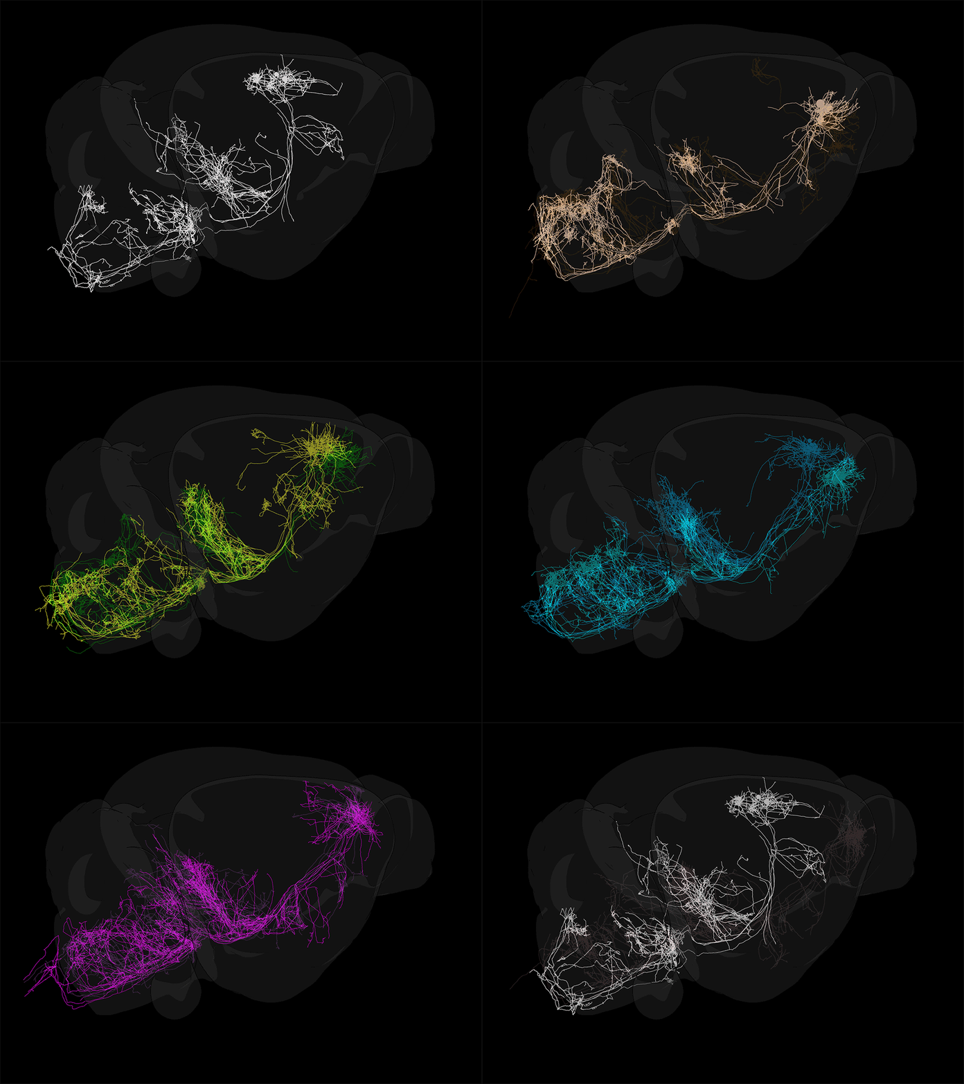 Single neurons in the motor cortex located in different subregions shown in different colors. (Image: Antonio Falasconi and Harsh Kanodia, Biozentrum, University of Basel)link.zoom
Mapping motor circuits
Antonio Falasconi and Harsh Kanodia from Arber’s team, together with their colleagues, analyzed 547 experiments tracing how signals travel from different parts of the motor cortex to other brain regions. This allowed them to compare connections with areas involved in movement, sensation and decision-making.
Areas with similar connection patterns clustered into 16 subregions arranged in three rows. Their organization followed two main directions. From back to front, the map shifted from areas more strongly connected to sensory regions, which process information such as touch and body position, toward areas linked to planning and decision-making. From side to side, the subregions corresponded broadly to different parts of the body, from the trunk and limbs to the face and mouth.
The team tested the map by tracing individual neurons and examining cell types across the cortex. Both approaches supported the same 16-subregion organization.
Motor cortex blueprint
The researchers also found that the primary and secondary motor cortex send coordinated signals to the brainstem and spinal cord, suggesting that the two regions may operate more in parallel than in the strict hierarchy often assumed.
“The most fascinating finding is the extremely high precision with which the motor cortical modules interact with the output regions and that the modules communicate to the rest of the cortex using the same wiring logic,” says Silvia Arber.
A reference map for brain research
The study used anatomical reference data and connectivity comparison tools from the Allen Institute’s Allen Institute’s Mouse Brain Atlas. Available through BrainGlobe, the new motor-cortex map provides a common framework for aligning research data. “Researchers interested in the cortex now have an accessible unified map to align their data to, and this will accelerate progress in the field,” says Falasconi.
The findings could also provide a basis for comparing motor-cortex organization across species, including humans. “Bringing together vast datasets describing the brain's wiring and its cellular makeup, we discovered a valuable and much more precise underlying blueprint of motor cortex organization,” says Kanodia.
Beyond movement research, the map could help scientists study conditions involving motor and frontal brain circuits, such as amyotrophic lateral sclerosis and frontotemporal dementia, by identifying which subregions and connections are especially vulnerable.
This text is based on a news of the Friedrich Miescher Institute for Biomedical Research.
Original publication
Antonio Falasconi et al.
Projection-defined modules reveal mouse motor cortex architecture
Cell (2026), doi: 10.1016/j.cell.2026.08.046