A switchboard with precision: How the brain licenses movements
Neurons deep in the brain not only help to initiate movement—they also actively suppress it, and with astonishing precision. This is the conclusion of a new study by researchers at the University of Basel and the Friedrich Miescher Institute for Biomedical Research (FMI), published in the journal Nature. The findings are especially relevant for better understanding neurological disorders such as Parkinson’s disease.
02 June 2025 | Livio Stöckli
Reaching for an apple or bringing a spoon to the mouth—these seemingly simple actions rely on highly complex processes in the brain. A key player in this orchestration is a deep-seated brain region known as the basal ganglia. For a long time, the output signal of the basal ganglia was thought to function mainly as a brake, suppressing unwanted behavior.
Researchers led by Professor Silvia Arber have now shown in mice that specific neurons in the basal ganglia make highly precise decisions about when to allow and when to actively stop a specific movement. Together, these dynamic signals license the timing of movement.
Basal ganglia: A central switchboard
These insights challenge the long-standing model of how the basal ganglia work. According to the traditional view, the basal ganglia control movement by continuously inhibiting motor centers in the brain, only briefly “releasing the brake” when a movement is allowed. “But this model falls far short in terms of complex movements, such as those involved in coordinated actions of the arms and hands,” explains Arber.

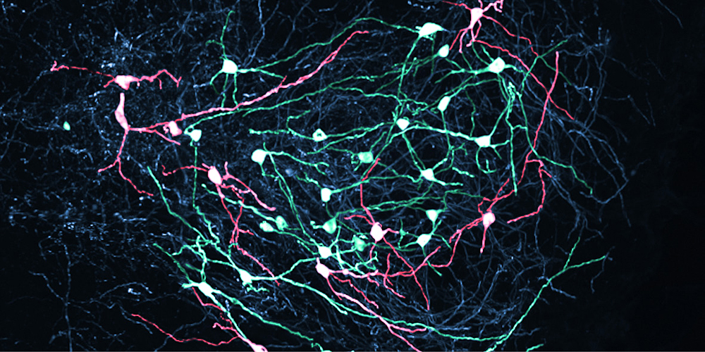
This study focuses on the so-called Substantia Nigra pars reticulata (SNr), the main output station of the basal ganglia, which sends signals to motor centers in the brainstem. The researchers made a surprising discovery: the neurons in this region don’t merely fire to inhibit movement. Instead, they display highly dynamic activity patterns —precisely timed to the movements being executed. During complex behaviors, SNr neurons switch multiple times between increased and decreased activity, each neuron with its specific dynamic pattern.
Thus, the output of the basal ganglia functions like a finely tuned system of traffic lights at a busy intersection: each light turns green or red for specific movements, depending on the action that is planned. In this way, complex behaviors can be built from individual movements, governed by the timing of these “go” and “stop” signals provided by SNr neurons.
Fine-grained movement control
To investigate these processes, two of Arber’s doctoral students recorded brain activity in mice as these used their hands to reach for food pellets. They found that individual SNr neurons responded very differently depending on the movement phase: when the arm reached, the hand grasped, or was retracted, specific neurons increased their activity while others paused. “It’s amazing how finely tuned these signals are,” Antonio Falasconi and Harsh Kanodia, the study’s lead authors, agree. “SNr neurons only pause their activity during very specific movements and increase it during select others.”
Using optogenetic techniques, the researchers then manipulated SNr neurons. They were able to show that activating these neurons blocked the behavior—a clear demonstration of their controlling role. Perhaps most strikingly, even the slightest changes in movement were accompanied by precise adjustments in SNr signaling. Downstream motor centers in the brainstem responded by sending signals back to the SNr.
So when the SNr “traffic light” turned green, the downstream neuron essentially presses the gas pedal, allowing the execution of a movement. This points to a highly specific, movement-based coding system—far more granular than just a general “go” or “stop” mechanism.
New avenues for treating movement disorders
The study offers a vivid picture of how the brain controls even the subtlest movements through a fine-tuned interplay of activation and inhibition—reshaping our understanding of motor control. This has important medical implications: in disorders like Parkinson’s or chorea, this delicate balance is disrupted, leading to hallmark symptoms such as difficulty initiating movement in Parkinson’s patients. “If we understand how the basal ganglia coordinate normal movement, we can develop more targeted treatments when this system goes out of balance,” explains lead researcher Arber.
Original publication
Silvia Arber, Antonio Falasconi, Harsh Kanodia et al.
Dynamic basal ganglia output signals license and suppress forelimb movements.
Nature (2025), doi: 10.1038/s41586-025-09066-z