How neuromuscular connections are maintained after nerve lesions
After nerve injury, the protein complex mTORC1 takes over an important function in skeletal muscle to maintain the neuromuscular junction, the synapse between the nerve and muscle fiber. Researchers at the University of Basel’s Biozentrum have now shown that the activation of mTORC1 must be tightly balanced for a proper response of the muscle to nerve injury. The study published in «Nature Communications» opens new insights into muscle weakness related to neuromuscular diseases or caused by ageing.
25 July 2019
The protein complex mTORC1 promotes muscle growth and is important for the self-cleaning process of the muscle cells. The role of mTORC1 in skeletal muscle fibers in response to nerve injury has so far not been studied in detail. New insights have been provided by the research team led by Markus Rüegg at the Biozentrum of the University of Basel.
Function of mTORC1
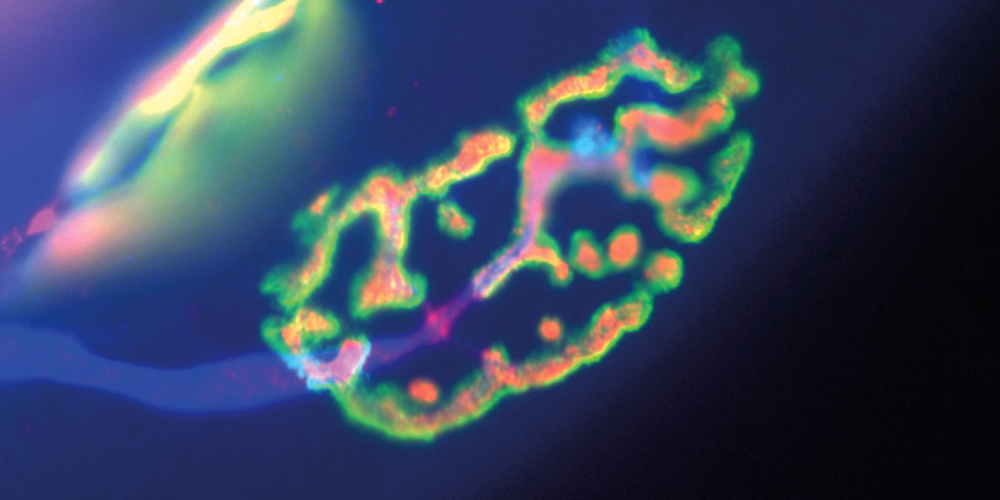
Nerves and muscles in our body are connected by specialized synapses, called neuromuscular junctions, which transmit signals from the nerve to muscle fibers. If innervation is lost or interrupted by the injury of the nerve, muscles cannot contract anymore. However, to prepare the muscle for re-innervation by the nerve, the muscle component of the neuromuscular junction, the motor endplate, is maintained. Rüegg’s research team has now more closely studied the function of mTORC1 and its upstream kinase PKB/Akt in the maintenance of the motor endplate after nerve injury using different mouse models.
The best known function of the PKB/Akt - mTORC1 signaling pathway is to promote muscle growth and the cellular self-cleaning process. «We have now been able to show that PKB/Akt and mTORC1 also play an important role in the maintenance of the neuromuscular endplate», explains Perrine Castets, first author of the study.
PKB/Akt - mTORC1 tightly balanced
After nerve damage, both PKB/Akt and mTORC1 get activated in muscle fibers. Rüegg‘s study demonstrates that mTORC1 should not be activated too strongly nor too little to ensure a proper response of the muscle. The underlying mechanism involves an mTORC1-dependent feedback onto the kinase PKB/Akt: «Should mTORC1 be too strongly activated, PKB/Akt is inhibited, resulting in the loss of the neuromuscular endplate. Balanced activation of both PKB/Akt and mTORC1 is required for the proper response of the muscle fiber», says Castets.
The newly described function of PKB/Akt and mTORC1 opens new perspective on how age-related muscle atrophy develops in humans. This is, likewise, induced through an alteration of neuromuscular endplates and possibly by over-activation of mTORC1. «Through this study, we now better understand the molecular mechanisms contributing to the maintenance of the neuromuscular junctions. Based on our results, we may be able to develop new approaches to potentially counteract age-related deficits and structural changes in order to better preserve the performance and functional capabilities of the muscles during ageing», says Rüegg.
Original source
Perrine Castets, Nathalie Rion, Marine Théodore, Denis Falcetta, Shuo Lin, Markus Reischl, Franziska Wild, Laurent Guérard, Christopher Eickhorst, Marielle Brockhoff, Maitea Guridi, Chikwendu Ibebunjo, Joseph Cruz, Michael Sinnreich, Rüdiger Rudolf, David J. Glass & Markus A. Rüegg
mTORC1 and PKB/Akt control the muscle response to denervation by regulating autophagy and HDAC4
Nature Communications (2019), doi: 10.1038/s41467-019-11227-4
Further information
- Prof. Dr. Markus A. Rüegg, University of Basel, Biozentrum, tel. +41 61 207 22 23, E-Mail: markus-a.ruegg@unibas.ch
- Heike Sacher, Universität Basel, Biozentrum, Communications, tel. +41 61 207 14 49, email: heike.sacher@unibas.ch