Collecting knowledge to combat eye diseases.
Text: Martin Hicklin
Researchers in Basel are investigating treatments for various eye diseases. Botond Roska is focusing on the retina through a series of experiments as well as exploring potential gene therapies.
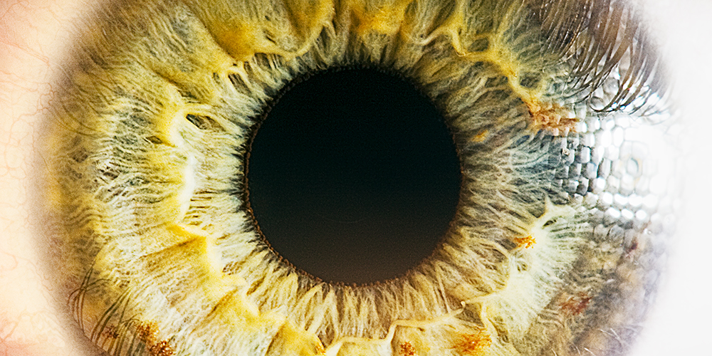
At the back of everyone’s retina, in the middle of the so-called “yellow spot” (macula lutea), lies a pit measuring just one and a half millimeters across. This fovea centralis is packed with 150,000 color-sensitive cones per square millimeter – and the signals they detect are converted, processed and relayed to the brain via the optical nerve. Damage to this site, which is responsible for high-acuity vision, makes daily tasks such as reading impossible, and there is nothing glasses can do to help.
“Those one and half millimeters are enough to keep half of the cerebrum busy,” says Botond Roska, Director of the Molecular Center at the Institute of Molecular and Clinical Ophthalmology Basel (IOB), emphasizing what a fascinating and miraculous role the retina plays. His mission is to understand what exactly goes on there with the aim of preventing blindness and giving affected individuals their eyesight back.
Born in Hungary in 1969, Roska already had the retina in mind when he came from Harvard to the Friedrich Miescher Institute in Basel in 2005. Meanwhile, his research group has conducted experiments to investigate the thin lining of the eye. For example, they have shown that the retina works like a biocomputer and that cells can adopt different roles. They also revealed the complex structure of the circuitry.
A new era in sight?
If Roska had not injured his hand in an accident, he might have become a famous cellist. But he was forced instead to swap his musical career for a degree in medicine. At Berkeley and Harvard, he would ultimately turn his attention to neurobiology and the retina in particular. Numerous awards – including, most recently, the Louis-Jeantet Prize for Medicine in spring 2019 – are a testament to his reputation as a leading retinal researcher. In Hendrik Scholl, Director of the Clinical Center at the IOB, Roska has found an ideal partner and friend.
Indeed, it appears that the two researchers and their colleagues have opened the door to a whole new era in ophthalmology. Progress seems to be coming in giant leaps right now. In 2018, for example, researchers succeeded for the first time in keeping a deceased donor’s retina in “incredibly good condition” for a whole day. Using fine electrodes, they were able to listen to the electrical murmur of the cells – a real turning point in this field of research. Now, they understand how to keep sections of a retina in working condition for up to 14 weeks and even how to restore a retina’s photosensitivity “optogenetically”, using smuggled-in genes. “This works remarkably well,” says Roska.
Access to viable retinas has also made it possible to examine the individual genes of the roughly 100 different cell types that work together there and to record which of them is being read and controlling the building of proteins. Known as “single-cell transcriptomics,” this process involves huge computing power and provides new insights into the development and function of the retina as an “image processor”. It also allows researchers to localize and map genetic defects.
Replacement of defective genes
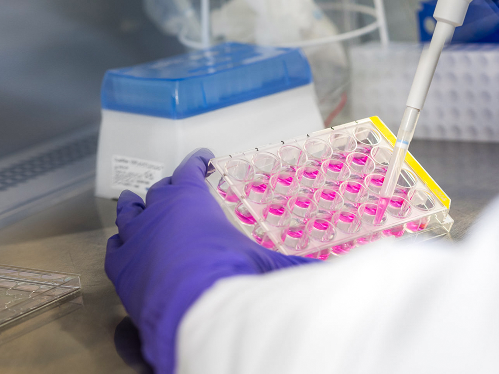
Roska’s enthusiasm shines through as he discusses these newly acquired abilities to grow artificial retinas and to carry out tests on “organoids” of this kind. Stem cells can now be made to differentiate in the retina in a process that uses skin cells as its starting material and coaxes them into converting back into pluripotent stem cells. In a suitable environment, these cells then grow into retinal organoids. These days, it is even possible to regrow a specific patient’s retina in the lab, with all its defects and peculiarities – providing an ideal basis for personalized medicine. Now, high-throughput techniques can also be used to test active substances on any number of organoids.
The best way to treat hereditary vision conditions, such as those that fall into the category of retinitis pigmentosa, is to replace the defective genes. This relies on gene vectors that can transport a working replacement into the defunct cells. The best candidates are “adeno-associated viruses” (AAVs), which only multiply in the presence of adenoviruses, do not cause disease, and barely trigger an immune response. “We were working with AAVs back when none of the major players were talking about them,” says Roska.
Today, the vectors are a highly sought-after commodity, and IOB recently led the publication of a library of 230 AAVs that are suitable for various cells and could be used for gene therapy. In collaboration with ETH Zurich’s Department of Biosystems Science and Engineering (D-BSSE) in Basel, the researchers developed a method whereby viruses are attached to magnetic nanoparticles, allowing them to be transported to the desired location more efficiently. In the case of Stargardt disease, in which only one letter of the genetic material is changed, there are plans for a precise “editorial” intervention.
Vision through the roof of the skull
One AAV vector from Roska’s lab is currently being used for the optogenetic treatment of advanced retinitis pigmentosa in a clinical trial in collaboration with GenSight Biologics in Paris. Running since 2018, this trial aims to restore some of the patients’ lost color sensitivity and to give the retina a genetic upgrade. Those receiving the treatment wear a pair of lightamplifying glasses. “There are 30 people working in my group, and they have thousands of different ideas,” says Roska. For example, researchers are considering how to make cells light-sensitive at an intermediate point on the way to the cerebrum in the event of a total failure of the optical nerve. This would allow visual information to be transported to the brain through the roof of the skull instead of the eye. It would also be conceivable to equip people with infrared sight.
“70 percent of my research focuses on basic principles, while the other 30 percent is clinically oriented,” says the researcher, adding that his work always looks at possible applications and how these can be translated into patient benefit. Growing insights may also pave the way for the treatment of widespread conditions such as age-related macular degeneration.
More articles in the current issue of UNI NOVA.


