Seeing, fast and slow.
Text: Angelika Jacobs
Signals from the peripheral fields of vision have a much longer path to the optic nerve than those from the center of the retina. How is it that we do not perceive a delay between our central and peripheral fields of vision?
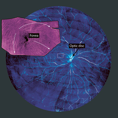
The human retina measures around five centimeters in diameter. All over the retina, light-sensing cells convert the light stimuli into electrical signals, which then travel via retinal nerve cells (more precisely, ganglion cells) to the optic nerve. The latter is a continuation of these ganglion cells, or rather their processes called axons, and connects the eye with the brain. It originates close to the center of the retina in the so-called “blind spot”. Signals from the peripheral fields of vision therefore have a much longer path to the optic nerve than those from the center of the retina. How is it that we do not perceive a delay between our central and peripheral fields of vision? Does the brain account for the temporal shift or does the synchronization already happen in the retina?
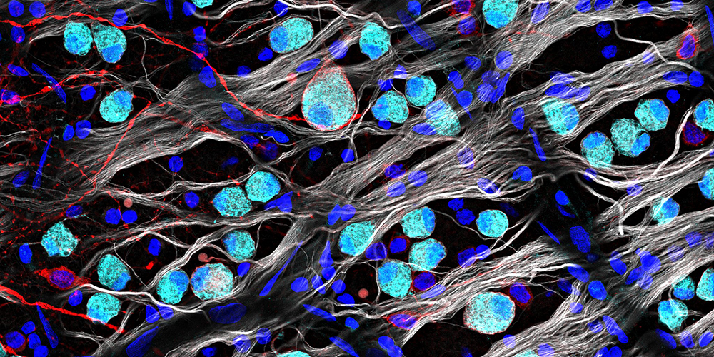
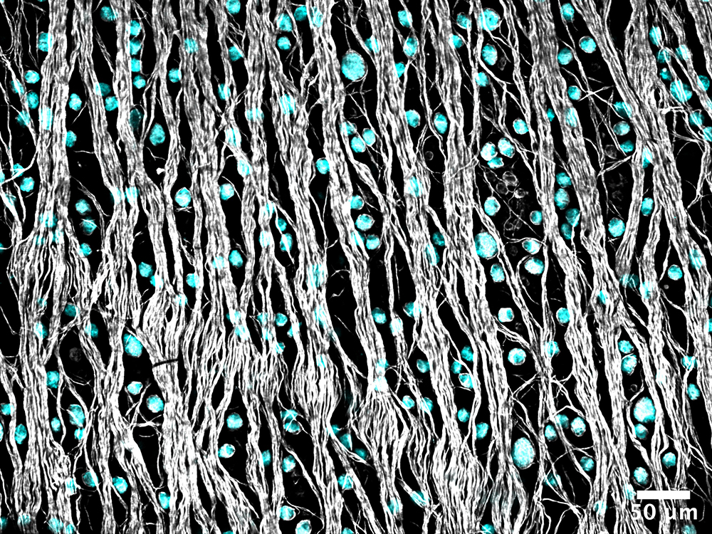
This question is being investigated by Felix Franke's research group at the Institute of Molecular and Clinical Ophthalmology Basel (IOB) in collaboration with colleagues at ETH Zurich. As part of her doctoral research, Annalisa Bucci examined human retinas from deceased persons who had donated their organs for research purposes. First, she stained the retinal cells to analyze the properties of the retinal ganglion cells and their extensions, the axons. Second, using an electrode-packed chip on which she placed a piece of retina, she was able to measure the speed of the signal traveling along the axons of individual ganglia.
On analyzing the traveling speeds, she found that signals from the periphery of the retina are indeed transported faster than those from the center. A similar compensatory effect comes into play when the axons curve around the fovea: This point of sharpest vision displays the highest density of light-sensing cells, and the axons of ganglion cells at the periphery take a detour to keep this area free of interfering structures. Yet, here the traveling speed of the signals also makes up for the extra distance.
The images were realized by Annalisa Bucci as part of her doctoral research.
More articles in the current issue of UNI NOVA.


