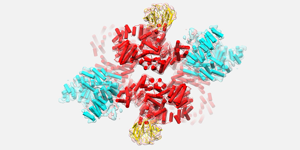
Architecture of cellular control center mTORC2 elucidated
The protein complex mTORC2 controls cellular lipid and carbohydrate metabolism. Researchers from the Biozentrum of the University of Basel and the ETH Zurich have now succeeded in deciphering the 3D structure of this important protein complex. The results have recently been published in “eLife”.
20 February 2018
The protein complex mTORC2 plays a crucial role in the regulation of cellular metabolism. It stimulates, for example, the production of lipids and fatty acids, but it also controls carbohydrate metabolism. In the past, studies have shown that mTORC2 promotes tumor growth due to increased lipid production and that it is involved in the development of diabetes.
The term mTORC2 derives from the fact that two such complexes exist in the cell. The two complexes, mTORC1 and mTORC2, are composed of different proteins, with the protein mTOR – “Target of Rapamycin” – as the central component.
Structure of mTORC2 solved
Using Nobel prize-winning cryo-electron microscopy, the scientists led by Prof. Timm Maier and Prof. Michael Hall from the Biozentrum of the University of Basel as well as Prof. Nenad Ban, ETH Zurich, have been able to visualize the three-dimensional structure of the mTORC2 protein complex in its assembled state.
“For the first time, we have obtained a 3D image of mTORC2 at intermediate resolution,” says Maier. “This picture not only reveals the basic shape of the complex, but also the interaction sites of accessory proteins with mTOR.” The two complexes, mTORC1 and mTORC2, are distinguished by accessory partner proteins of mTOR. This explains why the protein mTOR in its two complexes can perform distinct tasks in the cell.
The mTORC2 protein complex is one of the largest structures in the cell. It is about twenty times larger than a typical protein. But despite its size, it is still quite flexible and dynamic. “Its individual components move in different directions,” describes Maier. “Therefore, it was quite challenging to obtain a good quality 3D image of its structure.”
3D structure as basis for drug development
The 3D structure of a protein determines its function. Based on this structure, the researchers have made an important step towards investigating how mTORC2 recognizes its substrates and changes its shape as well as how the protein complex transmits signals in the cell.
“mTORC1 and mTORC2 signaling is linked to many diseases, but up to now there are no specific mTORC2 inhibitors available,” says Maier. "The structural insights may facilitate the search for such drugs.”
Original article
Edward Stuttfeld, Christopher H.S. Aylett, Stefan Imseng, Daniel Boehringer, Alain Scaiola, Evelyn Sauer, Michael N. Hall, Timm Maier, Nenad Ban
Architecture of the human mTORC2 core complex
eLife (2018), doi: 10.7554/eLife.33101
Further information
- Prof. Dr. Timm Maier, University of Basel, Biozentrum, tel. +41 61 207 21 76, email: timm.maier@unibas.ch
- Dr. Katrin Bühler, University of Basel, Communications Biozentrum, tel. +41 61 207 09 74, email: katrin.buehler@unibas.ch


