Researchers build bone marrow model entirely from human cells
Our body’s “blood factory” consists of specialized tissue made up of bone cells, blood vessels, nerves and other cell types. Now, researchers have succeeded for the first time in recreating this cellular complexity in the laboratory using only human cells. The novel system could reduce the need for animal experiments for many applications.
18 November 2025 | Angelika Jacobs
The bone marrow usually works quietly in the background. It only comes into focus when something goes wrong, such as in blood cancers. In these cases, understanding exactly how blood production in our body works, and how this process fails, becomes critical.
Typically, bone marrow research relies heavily on animal models and oversimplified cell cultures in the laboratory. Now, researchers from the Department of Biomedicine at the University of Basel and University Hospital Basel have developed a realistic model of the bone marrow engineered entirely from human cells. This model may become a valuable tool not only for blood cancer research, but also for drug testing and potentially for personalized therapies, as reported by a team of researchers led by Professor Ivan Martin and Dr Andrés García García in the journal Cell Stem Cell.
Bone marrow architecture and cell diversity
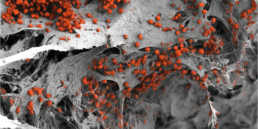
Bone marrow is not uniform, it is made up of several specialized microenvironments, also known as “niches”. One niche that is particularly important for blood formation and is related to blood cancer’s resistance to therapies is located close to the bone surface. The so-called endosteal niche includes blood vessels, bone cells, nerves and immune cells. Until now, there had been no human bone marrow model that included all these cellular components.
The research team has now successfully created such a model. The basis for this complex tissue was an artificial bone structure made from hydroxyapatite, a natural component of bones and teeth. The team used human cells reprogrammed into pluripotent stem cells using molecular biology methods. These artificially developed stem cells can produce different specialized cell types depending on the signals they receive in their environment.
The researchers integrated these stem cells into the artificial bone structure and guided them through specific differentiation processes to produce a wide range of bone marrow cell types in a reproducible and controlled way. Their subsequent analysis confirmed that this three-dimensional construct closely resembles the human endosteal niche and is larger than previous systems, measuring eight millimeters in diameter and four millimeters in thickness. The described model allowed the researchers to sustain human blood formation in the laboratory for weeks.
A step toward replacing some animal experiments
“We have learned a great deal about how bone marrow works from mouse studies,” says Ivan Martin. “However, our model brings us closer to the biology of the human organism. It could serve as a complement to many animal experiments in the study of blood formation in both healthy and diseased conditions”. This aligns with the university’s efforts to replace, reduce, and refine animal experiments whenever possible.
The system could also be used in drug development in the future. “However, for this specific purpose, the size of our bone marrow model might be too large,” explains Andrés García García. In order to test multiple compounds and doses in parallel, the model would need to be miniaturized.
In the long term, the use of the model in the definition of personalized treatments for blood cancers is also conceivable, with individual bone marrow models being generated using patients’ cells in order to test different therapies and select the most effective one for each patient. However, the researchers acknowledge that this will also require further development.
Original publication
Qing Li et al.
Macro-scale, scaffold-assisted model of the human bone marrow endosteal niche using hiPSC-vascularized osteoblastic organoids.
Cell Stem Cell (2025), doi: 10.1016/j.stem.2025.10.009